-
entries
0 -
comments
0 -
views
287
About this blog
Do tattoos cause cancer?
Can tattoos cause cancer? Researchers have been exploring this problem for many years. Although there is no direct evidence that tattoos can cause cancer, people always regard tattoos as an established fact. When it comes to cancer, black ink is undoubtedly an unavoidable factor. As the most commonly used color for tattoos, black ink contains high levels of benzo(a)re, which is classified as a carcinogen by the International Agency for Research on Cancer (IARC). At the same time, blackout tattoos have sprung up in the past period of time. This new thermal trend may be particularly dangerous because it requires thick black solid ink to cover most of the individual’s body. In addition to worrying about carcinogens in ink, individuals are also concerned about the way these tattoos cover the body. Changes in skin pigmentation are one of the earliest signs of skin cancer, especially melanoma. When the body is “blackened” by tattoo ink, individuals may not immediately notice these changes. Therefore, never place the tattoo on the original mole, birthmark or other skin discoloration or abnormality.
Another reason for concern is what happens after a certain period of time. Over time, tattoos tend to fade and lose pigmentation. This is especially true for black tattoos that tend to break down faster. When a tattoo begins to fade and lose pigment, it produces many carcinogenic compounds.
There is no doubt that there are various links between tattoos and cancer. But some recent scientific studies have shown that tattoos are not only one of the carcinogenic factors, but also help to find cancer.
Tattoo help detect cancer
As we all know, many cancers are usually found only in the advanced stages. At this time, treatment is very difficult and the possibility of cure is very small.
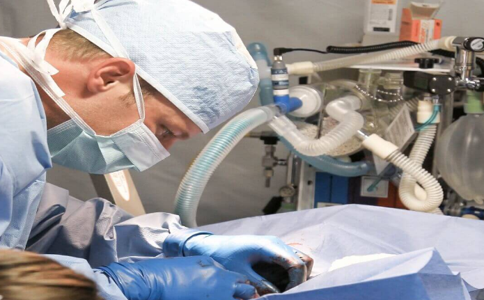
As early as 2018, Professor Martin Fusenger from the Department of Biological Systems Science and Engineering at ETH Zurich has developed another type of “tattoo” that can accurately detect cells that may exist in the early stages of cancer. The researchers conducted a feasibility study to test the effectiveness and accuracy of the implant in predicting skin cancer in mice and pigs. The results of their research currently seem very promising, and the research has been published in the journal Science Translational Medicine.
And now, according to the latest research from the Viterbi Department of Biomedical Engineering at the University of Southern California, the humble ink on the needles of tattooists may be the key to improving cancer detection rates. Cristina Zavaleta and her team have recently developed new imaging contrast agents using common dyes such as tattoo dyes and food dyes. When these dyes are attached to nanoparticles, they can illuminate cancer, allowing medical professionals to better distinguish cancer cells from normal neighboring cells. This work has been published in “Biomaterials Science”.
Many cancers can only be found at a very high stage, and treatment is ineffective at this time, which means that it is difficult for patients to recover. Early detection is essential for patients to achieve the best cancer outcome. This disease affects more than 38% of Americans at some point in their lives.
Fussenegger explained: “Early diagnosis can significantly increase the chances of survival. For example, if breast cancer can be detected early, the cure rate is 98%. However, if the diagnosis is too late, only a quarter of women can survive. Now Tumors can cause health problems only when people go to the doctor. Unfortunately, it’s too late.”
Zavaleta has the same explanation for this: “For example, if the problem is colon cancer, it can be found through endoscopy.” “But the endoscope is actually just a flashlight at the end of a stick, so it can only provide information about the structure of the colon. -You can see polyps and know that a biopsy is needed.” “But if we can provide imaging tools to help doctors see if a particular polyp is cancerous or benign, maybe they don’t even need to take it.” She said
Professor Fussenegger and his team believe that the special skin implants they designed may greatly improve this situation in the future. They call it a “biomedical tattoo.” “Biomedical tattoos” look like brown moles. If they are “ignited”, it indicates early symptoms of cancer. They use biologically sensitive ink to monitor health. If the composition of human tissue fluid changes, the ink will change color.
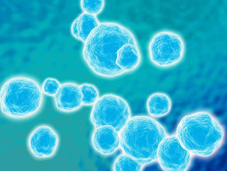
The Zavaleta team believes that contrast materials, when injected into patients, can make imaging functions such as MRI and CT more sensitive and specific so that medical professionals can accurately diagnose and allow surgeons to identify tumors. The exact edge. Luminous nanoparticles move in blood vessels to find cancer. Coloring dyes are incorporated into nanoparticles to obtain more sensitive imaging contrast when identifying cancer cells.
Their biomedical tattoos will be able to identify the four most common types of cancer, which are usually found late, including breast, lung, prostate and colon cancer.
Working principle
In the early stages of cancer, blood calcium levels rise, a phenomenon called “hypercalcemia”. Studies report that 30% of patients diagnosed with cancer have elevated blood calcium levels.
The implant consists of a series of “genetic components” incorporated into somatic cells. Once implanted under the skin, the implant can monitor the calcium level in the blood.
If the calcium level in the blood rises abnormally, melanin will fill these genetically modified cells, making them look like brown moles. Therefore, the “implanted person” can detect the symptoms of early cancer.
Professor Fussenegger said: “If a brown mole appears, the implanted person can go to the doctor for further evaluation. The appearance of a brown mole does not mean that death will soon. On the contrary, the implanted person should regard it as an early signal, You should see a doctor to check your health.”
As Professor Fussenegger pointed out, implants are mainly used for self-monitoring, so this is a very cost-effective method.
However, if a person does not want to feel pressured because this ignited artificial “mole” represents a potential cancer, then they have another option.
Professor Fussenegger and colleagues have also developed an alternative implant in which hypercalcemia will only cause markings that are visible under a special red light, similar to “invisible ink.” Fisengege said: “This means that the implanted person needs to be checked regularly with the doctor.”
According to the vision of the Zavaleta team, contrast materials which when injected into patients, allow for imaging such as MRI and CT to function with better sensitivity and specificity, enabling medical professionals to diagnose with accuracy, and for surgeons to identify the exact margins of tumors
In order to achieve this goal, the research team discovered a unique source of optical contrast agents from the household coloring dyes and pigments we often encounter. These “optical inks” can be attached to cancer-targeted nanoparticles to improve cancer detection and localization.
Dyes and pigments are found in common colorants that have been approved by the U.S. Food and Drug Administration (FDA), and the research team hopes that this will make them easier and safer to implement in imaging practices.
For Zavaleta, the inspiration came from an unusual place an animation class attended by Pixar animators in Emeryville, California, where the famous studio is located. Zavaleta loves art and animation. She said that she is very interested in the ink paintings that artists bring to class.
Zavaleta said: “I’m thinking about how these real high-pigment paints (such as gouache watercolor paints) can become bright in an unprecedented way, and I want to know if they have interesting optical properties.”
This idea led her to find a tattooist in nearby San Francisco, Adam Sky, another artisan who uses bright dyes.
Zavaleta said: “I remember I brought a 96-well plate, and he sprayed tattoo ink in each well.” “Then I took the ink to our Raman scanner (used to detect targeted tumors sensitively). Nanoparticles), found these very amazing spectral fingerprints, which can be used to barcode nanoparticles. This is so cool.”
One of the safety challenges of using nanoparticles for imaging is that these nanoparticles usually stay longer in organs such as the liver and spleen, which are responsible for breaking down the nanoparticles. Due to these safety issues, it is important to consider biodegradable nanomaterials. Currently, the number of optical contrast agents approved for clinical use is limited.
With this in mind, Zavaleta’s team considered common food dyes that can be used to decorate nanoparticles, such as those found in skittles and colored candies in M&M. The FDA believes that these brightly colored foods consumed by humans are safe for human consumption.
Zavaleta said: “We think, let’s take a look at some FDA-approved drugs, cosmetics and food dyes to see what optical properties are in them.” “So, we finally found that many of these FDA-approved dyes have interesting optical properties and are available. For imaging.”
The team has developed a nanoparticle that will carry these pigment-rich imaging agents as a “payload.” Zavaleta said that these particles have a specific size that allows them to passively penetrate the tumor area, but they can also be retained due to their size.
The progress of the experiment
The tests so far have confirmed that this implant can be used as a reliable diagnostic aid, but there are still some shortcomings. The main problem is that its shelf life is not long, so it needs to be updated frequently and repeatedly.
Professor Fussenegger pointed out: “The encapsulated living cells can survive for about a year. After that, they must be inactivated and replaced.”
Another problem is that the implant is still in the early stages of prototype and needs further research before it can be used for human testing. The road to applying such biomedical tattoos to humans is still very difficult and long.
Professor Fussenegger explained: “Continuous development and clinical trials are very difficult and costly. As a research team, it is difficult for us to afford it.” He admitted that the entire research process may take more than ten years to complete. But he added that it is worth the wait and hard work because this is a concept that can be adopted. It can help early diagnosis of many diseases, from various neurodegenerative diseases to hormone abnormalities.
The Zavaleta team believes that most imaging contrast agents in clinical use today are small molecule dyes.
Zavaleta said: “With small molecules, you may be able to see them initially accumulate in the tumor area, but you have to be faster until they finally leave the tumor area and excreted.” “Our nanoparticles happen to be small enough to penetrate, But at the same time it is large enough to be retained in the tumor. This is what we call enhanced permeability and retention.”
Zavaleta said: “With small molecules, you may be able to see them initially accumulate in the tumor area, but you have to be faster until they finally leave the tumor area and excreted.” “Our nanoparticles happen to be small enough to penetrate, But at the same time it is large enough to be retained in the tumor. This is what we call enhanced permeability and retention.”
Compared with previous small molecule imaging agents, nanoparticles can also be “modified” with larger dye payloads. The research team has shown that nanoparticles lead to brighter signals and obvious positioning of nanoparticles in tumors under fluorescence imaging.
Zavaleta said: “If you encapsulate a bunch of dyes in nanoparticles, you will be able to see it better because it will be brighter.” “It’s like using a bag of dyes instead of just using one dye.”
Future development
Today is different from the past. The standards and requirements of the tattoo industry are becoming stricter. All tattoo equipment must undergo various inspections and can only be sold and used after being qualified. Although our tattoo ink has not yet reached the effect of detecting cancer, we will continue to pay attention to this experiment. Once the ink can be put into use, our website will be sold as soon as possible. At present, the tattoo ink sold on our website has reached drinking level and is completely harmless. Please rest assured to buy!